
Dermatology/Mole Mapping
98.4% of Melanomas are curable if found early
One of the most important things after being diagnosed with melanoma is learning its stage (extent of the melanoma at the initial/primary site and extent, if any, that melanoma has spread elsewhere in your body). Knowing the stage of melanoma helps to determine your initial prognosis and helps your clinical team identify the best treatment plan for you.
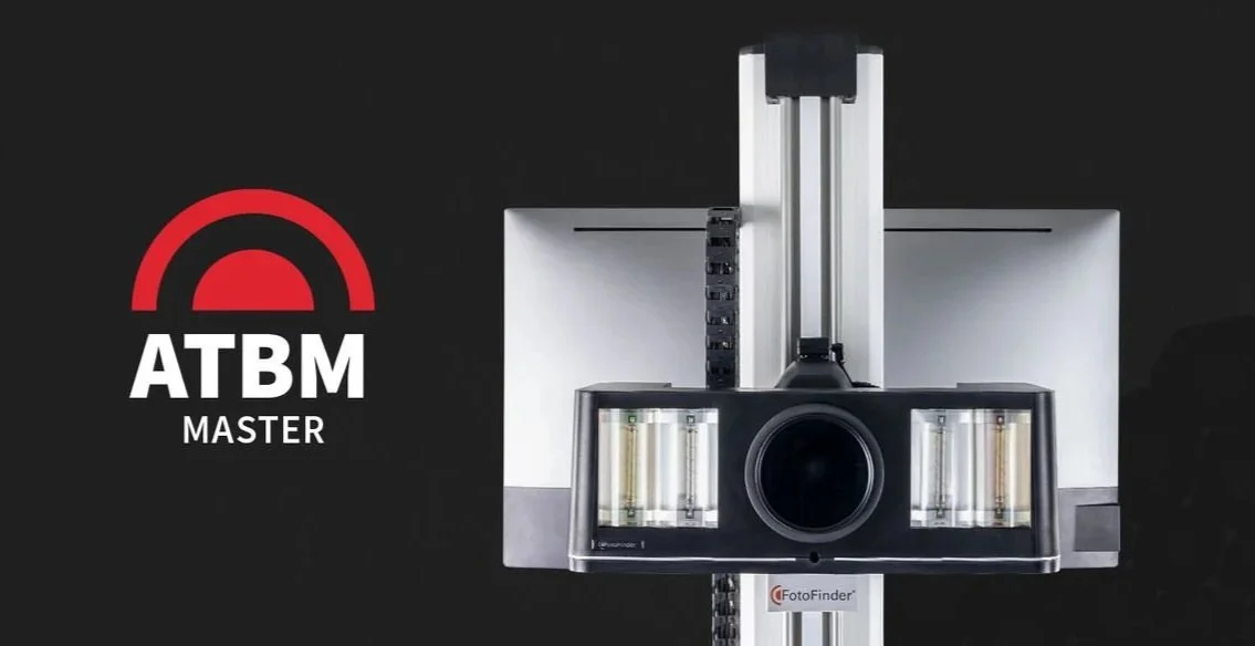
Introducing Fotofinder
Integrated Health Clinic is thrilled to now provide the best-in-class Advanced Total Body Mapping & Dermoscopy for skin lesions.
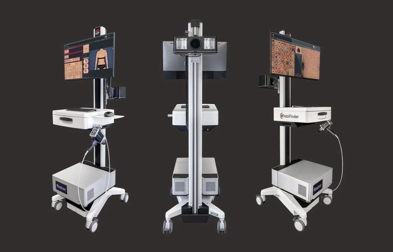
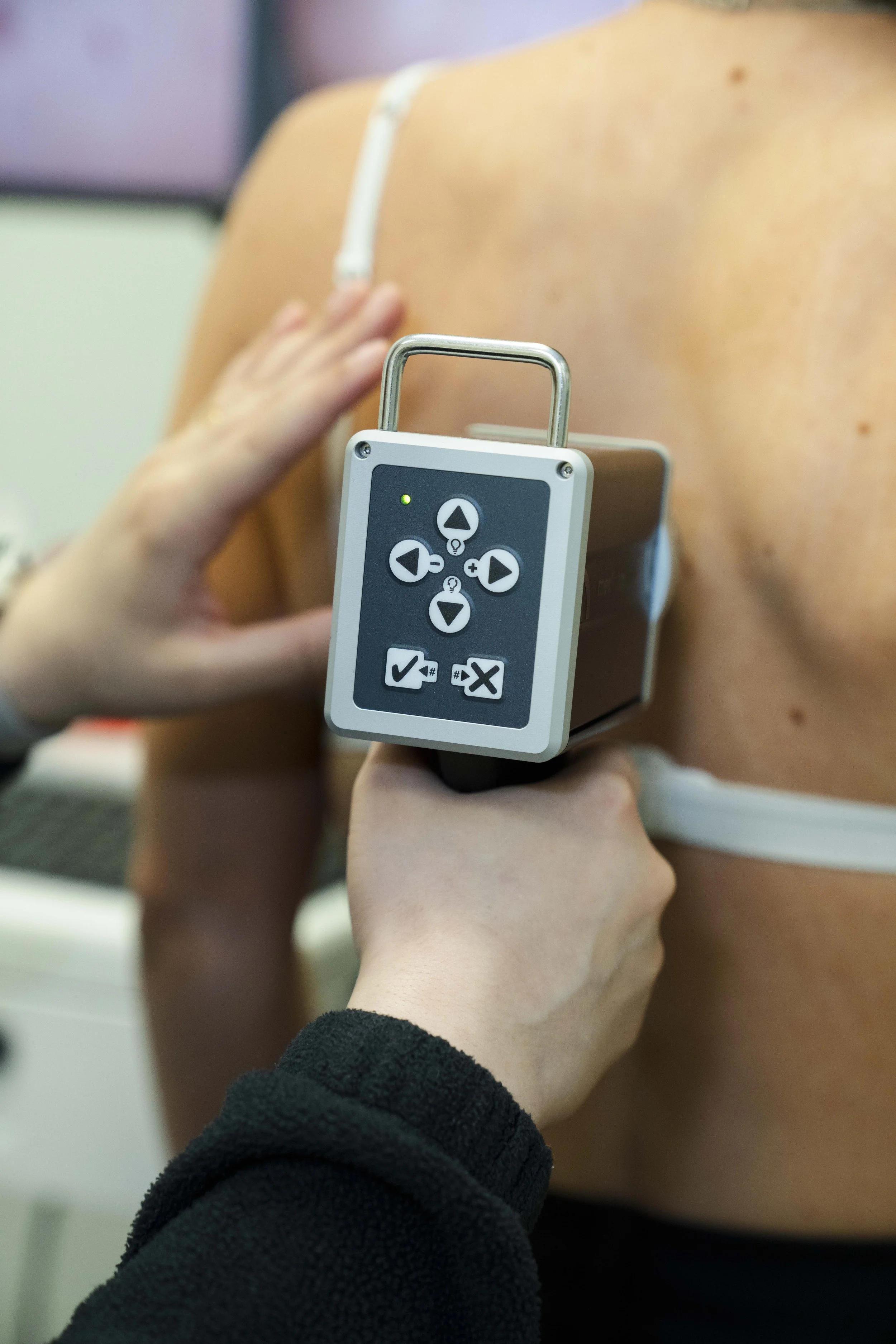
The rates of skin cancer, including malignant melanoma, are on the rise. The fact remains that if found early, skin cancers are largely curable. The key to early diagnosis FotoFinder ATBM MASTER is an automated Full Body Photography system that captures detailed photos of your entire skin.
The high-resolution camera is connected to a computer that transfers all photos directly to the doctor’s database, giving the physician the ability to not only find skin cancers, but then to precisely compare photos from your initial visit to identify any new lesions or changes to existing lesions. In a second step highly magnified images, or dermoscopy, of identified concerning skin lesions are captured to track any changes over time. The photography session takes only a few minutes and is completely painless.
What is Dermoscopy?
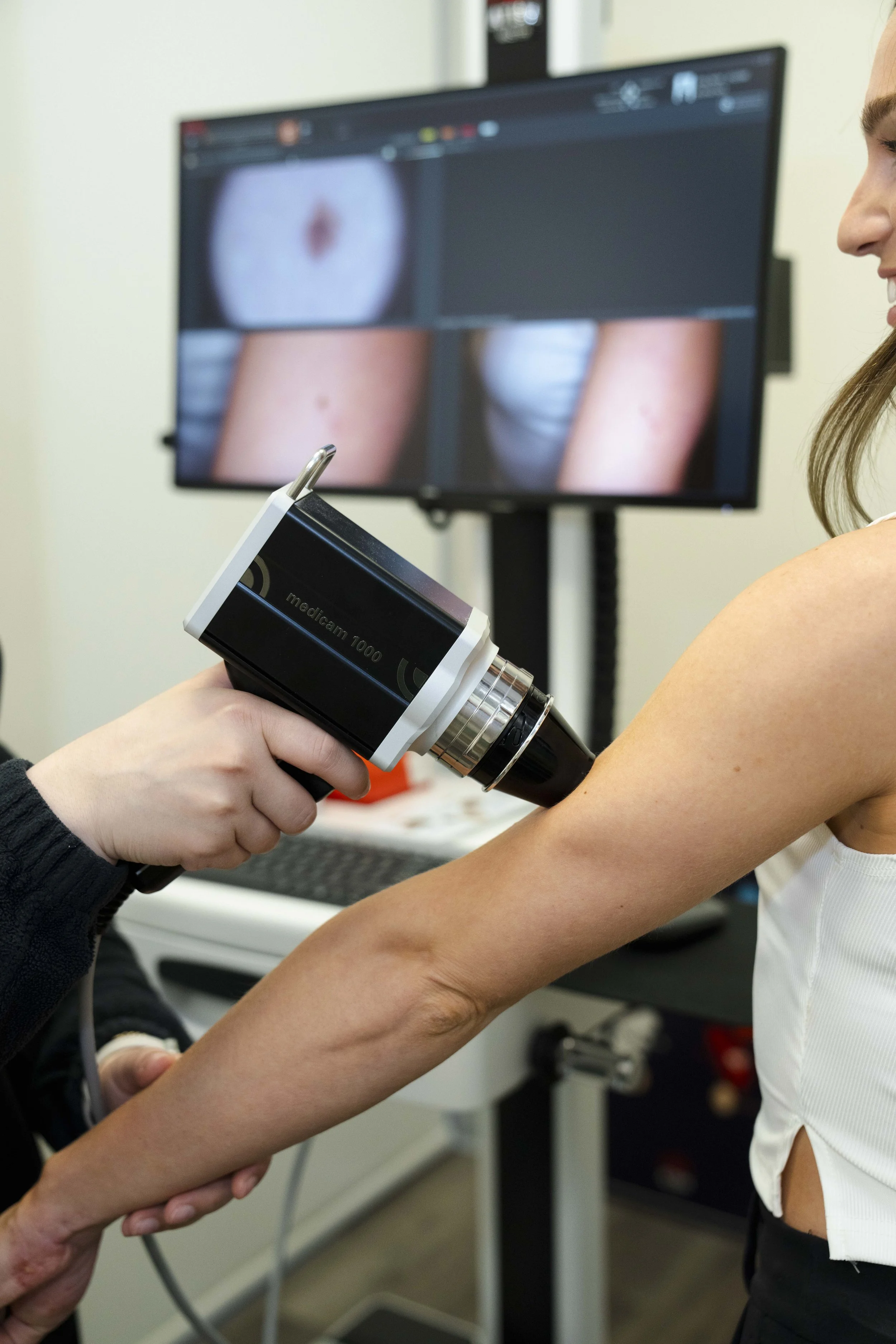
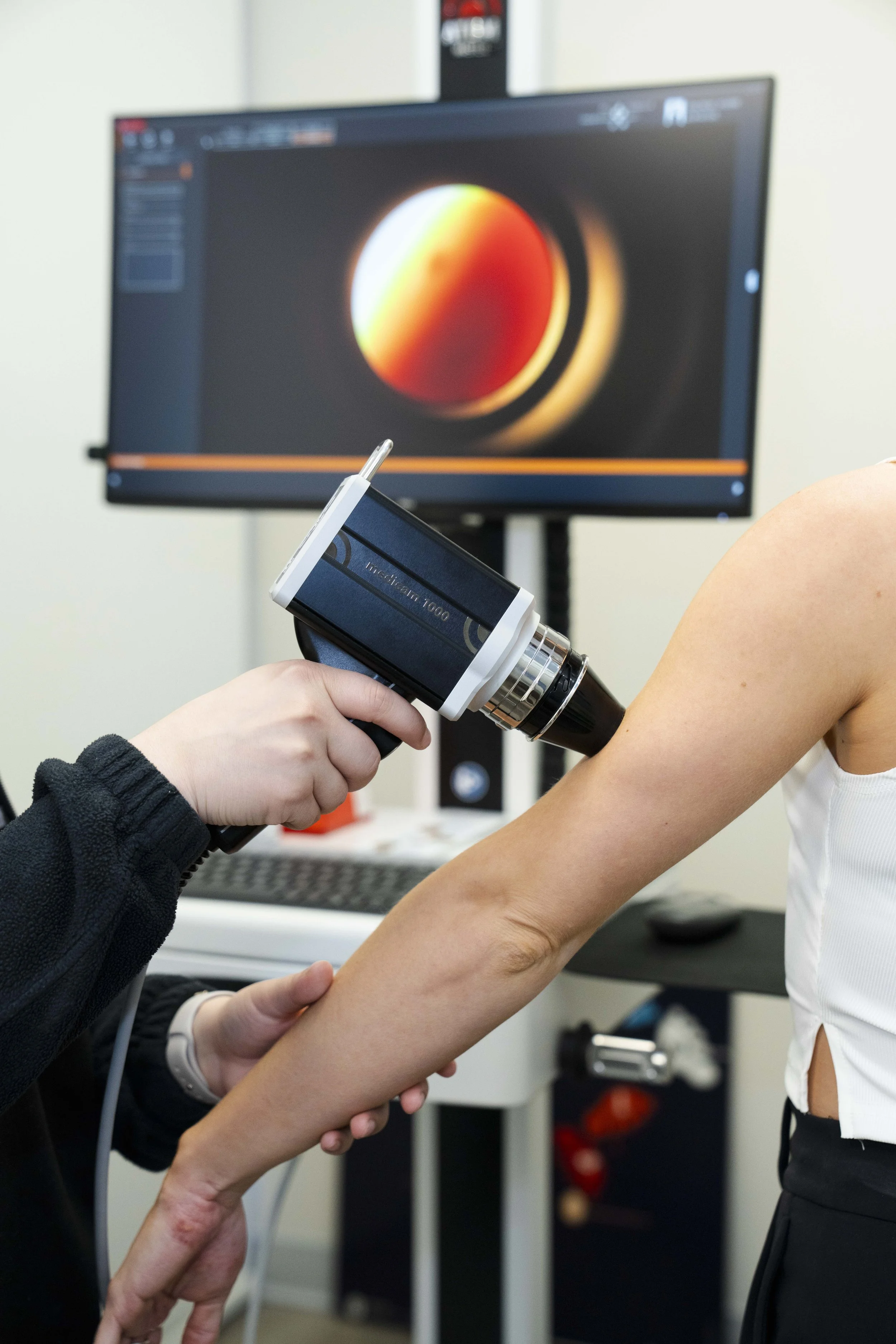
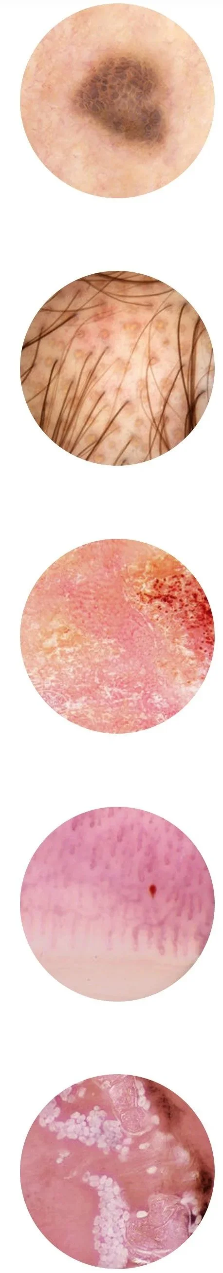
The Medicam 1000 HD camera provides a visual perspective of skin lesions never before seen, allowing your doctor to find, assess and monitor skin lesions with tremendous precision (20x-120x magnification). The Medicam 1000s offers spectacular sharpness in video dermoscopy and surpasses its predecessors and rivals in image quality and performance.
Typical Process
An easy and reproducible sequence of images are taken from head to toe within just minutes. The doctor will review these ATBM images and then perform a complete skin exam, taking more detailed dermoscopic images as needed to complete the examination. Concerning lesions may then be removed for pathological evaluation, providing a diagnosis and the next steps for care. Sometimes that means a repeat dermoscopic examination in 3 months to look for evolution, a biopsy, or to repeat the ATBM process annually.
Potential in-office procedures available at IHC
Punch Biopsy of skin lesion
Excisional Biopsy of skin lesion
Shave Biopsy of skin lesion
Angioma Removal with electrosurgery
Wart Removal with electrosurgery
Skin tag Removal with electrosurgery
Seborrheic keratosis Removal with electrosurgery
Telangiectasias Removal with electrosurgery
Pyogenic granuloma Removal with electrosurgery & curettage
How ATBM (Mole Mapping) & Dermoscopy Can Help
Allows observer to concentrate on lesion and formulate a logical differential diagnosis
Helps differentiate melanocytic from non-melanocytic lesions
Helps differentiate benign from malignant lesions
Improves diagnostic accuracy
Increases the observer’s confidence in their clinical diagnosis
Confirms naked eye diagnosis (clinical-dermoscopy correlation)
Improves malignant to benign biopsy ratio (avoids unnecessary biopsies)
Helps isolate suspicious foci within lesions – directing pathology
Helps more precisely define borders of some lesions for improved pre-surgical margin mapping
Helps the surveillance of patients with many nevi (moles)
Helps reassure patients
Reference: Benvenuto C, Marghoob AA. Ten reasons why dermoscopy is beneficial for the evaluation of skin lesions. Expert Rev Dermatol. 2006; 1(3):1-6.